


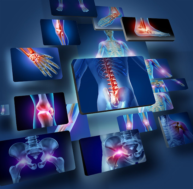
Medical imaging scans have revolutionized modern healthcare by allowing doctors to see inside the body without surgery. These technologies play a crucial role in early diagnosis, monitoring, and treatment planning for many conditions.
X-Rays: Fast Imaging for Bones and Dense Tissue
X-rays are one of the oldest and most commonly used imaging methods. They work by passing small amounts of radiation through the body to create images of bones and dense tissues.
- Ideal for detecting fractures and bone conditions
- Commonly used in dental and chest imaging
- Quick and widely available
However, X-rays use ionizing radiation, which may carry a small risk when used frequently. They are generally avoided during pregnancy unless absolutely necessary.
Ultrasound: Safe and Radiation-Free Imaging
Ultrasound, also known as sonography, uses high-frequency sound waves to create images of internal organs and tissues.
- Commonly used during pregnancy
- Helps diagnose abdominal and organ conditions
- Safe because it does not use radiation
Ultrasound is often recommended for examining soft tissues and is frequently used alongside procedures like abdominal scans.
MRI: Detailed Imaging of Soft Tissues
Magnetic Resonance Imaging (MRI) uses strong magnetic fields and radio waves to produce detailed images of internal structures, especially soft tissues.
- Useful for brain, spine, and joint imaging
- Helps detect tumors and neurological conditions
- Does not use ionizing radiation
According to the Cleveland Clinic explanation of MRI scans, this technology provides highly detailed images that are essential for diagnosing complex conditions.
However, MRI scans require patients to remain still and may not be suitable for those with certain metal implants.
CT Scans: Cross-Sectional Imaging
Computed Tomography (CT) scans combine multiple X-ray images to create detailed cross-sectional views of the body.
- Provides detailed images of organs, bones, and blood vessels
- Useful in emergencies such as trauma or stroke
- Fast and highly accurate
CT scans expose patients to more radiation than standard X-rays, so they are typically used only when necessary.
Nuclear Imaging: Advanced Functional Imaging
Nuclear imaging involves the use of small amounts of radioactive material to visualize how organs function.
- Used to detect cancers and heart conditions
- Provides functional information, not just structure
- Requires time for the tracer to circulate in the body
This method offers valuable insights but is generally reserved for more complex diagnostic needs.
Risks and Safety Considerations
While medical imaging is generally safe, each type carries its own risks:
- Radiation exposure from X-rays and CT scans
- Magnetic interference in MRI scans
- Allergic reactions to contrast dyes
Doctors carefully weigh the benefits and risks before recommending any imaging test.
When Are Imaging Scans Necessary?
Imaging tests are often used when symptoms cannot be explained through physical examination alone. For example, persistent pain may require further investigation similar to cases discussed in back pain diagnosis.
They are also essential for monitoring treatment progress and guiding surgical procedures.
Conclusion
Medical imaging scans provide valuable insights into the human body, helping doctors diagnose and treat a wide range of conditions. From X-rays to advanced nuclear imaging, each technique serves a specific purpose.
Understanding how these scans work can help patients feel more informed and confident when undergoing diagnostic procedures.
Frequently Asked Questions
Ultrasound is generally considered the safest because it does not use radiation.
CT scans involve higher radiation exposure than X-rays, but they are safe when used appropriately.
MRI is excellent for soft tissue imaging but may not be ideal for all types of conditions.
They help diagnose conditions that cannot be identified through physical examination alone.
No, most imaging scans are painless, though some may require contrast injections or staying still for extended periods.
