


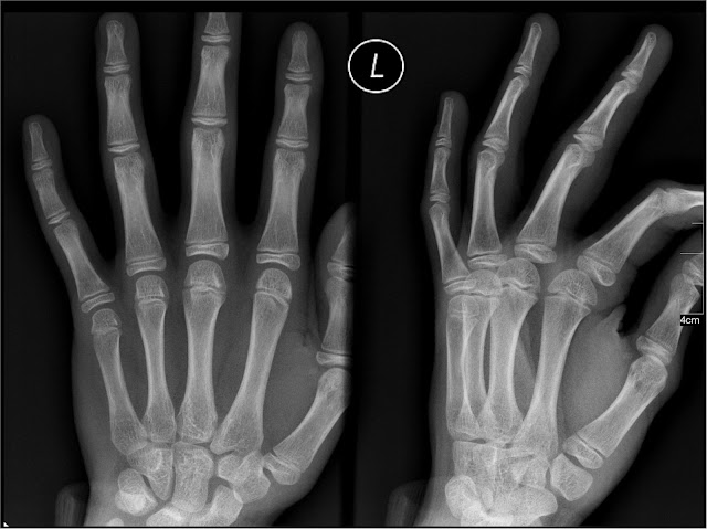
X-ray imaging is one of the oldest and most widely used medical diagnostic tools. First discovered in 1895 by Wilhelm Röntgen, X-rays revolutionized medicine by allowing doctors to see inside the human body without surgery. Today, despite advances in imaging technologies, X-rays remain essential in diagnosing a wide range of conditions.
What Are Medical X-Rays?
X-rays are a form of electromagnetic radiation, similar to visible light but with much shorter wavelengths. This allows them to penetrate the body and produce images of internal structures.
In medical settings, X-rays are generated using a specialized device called an X-ray tube. When a high-voltage electrical current passes through the tube, it produces radiation that travels through the body and is captured to form an image.
Different tissues absorb X-rays differently:
- Bone: absorbs more radiation and appears white
- Soft tissue: appears in shades of gray
- Air: absorbs very little radiation and appears black
Radiography Equipment and How It Works
X-ray machines consist of a radiation source and a detector. When X-rays pass through the body, they are partially absorbed depending on the density of tissues. The remaining rays hit a detector to create an image.
This process is widely used in diagnosing injuries such as fractures or conditions like spinal disc problems and chest infections.
The Shift to Digital X-Ray Imaging
Traditionally, X-ray images were captured on photographic film, which required chemical processing and physical storage. Modern healthcare has largely transitioned to digital X-ray systems.
Digital imaging offers several advantages:
- Instant image viewing
- Reduced need for physical storage
- Enhanced image quality and contrast
- Easier sharing between healthcare providers
These improvements have made diagnostics faster and more efficient, especially in emergency settings and when monitoring recovery after procedures like back surgery recovery.
Common Uses of Medical X-Rays
X-rays are primarily used to evaluate bones, but they also help visualize certain soft tissues and organs. They are essential in diagnosing conditions such as fractures, infections, and tumors.
Common applications include:
- Detecting bone fractures and joint issues
- Diagnosing lung conditions like pneumonia
- Identifying kidney stones
- Monitoring digestive system activity
X-rays also play a role in specialized imaging techniques:
- Dental X-rays: detect cavities and tooth problems early
- Mammography: screens for breast cancer
- Fluoroscopy: provides real-time moving images for procedures
For a broader overview of imaging techniques, you can explore medical imaging scans.
Are Medical X-Rays Safe?
X-rays involve exposure to low levels of radiation. While any radiation carries some risk, the dose from a standard X-ray is minimal.
According to guidelines on medical radiation safety, the benefits of accurate diagnosis typically outweigh the small risks associated with X-ray exposure.
However, certain groups require extra caution:
- Pregnant women
- Young children
- Patients requiring repeated imaging
Doctors carefully evaluate the necessity of each scan to minimize unnecessary exposure.
Other Medical Uses of X-Rays
Beyond diagnostic imaging, X-rays are also used in therapeutic and advanced imaging applications:
- Radiotherapy: uses high-energy radiation to treat cancer
- CT scans: combine multiple X-ray images to create detailed cross-sectional views
While CT scans provide more detailed images, they expose patients to higher radiation levels compared to standard X-rays.
Conclusion
Medical X-rays have evolved significantly since their discovery, transitioning from film-based imaging to advanced digital systems. Despite newer technologies, they remain a cornerstone of modern diagnostics due to their speed, accessibility, and effectiveness.
Understanding how X-rays work and their safety profile helps patients feel more confident when undergoing imaging procedures as part of their medical care.
Frequently Asked Questions
X-rays are primarily used to visualize internal structures of the body, especially bones, to diagnose injuries and diseases.
Yes, digital X-rays provide faster results, improved image quality, and easier storage and sharing compared to traditional film-based imaging.
The radiation dose from a standard X-ray is very low and generally considered safe when used appropriately.
While X-rays are best for bones, they can also help identify certain soft tissue issues, especially in the lungs and digestive system.
Pregnant women and young children should avoid unnecessary X-rays unless medically required, due to increased sensitivity to radiation.
