Medical X-rays – Radiological Imaging From Film to Digital X-rays

medical x-rays

X-ray pictures were the earliest medical imaging technique. The first X-ray film of the human body was taken in 1895 by the discoverer of X-rays Wilhelm Röntgen, showing his wife’s left hand with her wedding ring. Despite the development of newer technologies such computed tomography (CT), magnetic resonance imaging and ultrasonography, X-rays remain important for the diagnosis of many disorders.

Radiography Equipment

X-rays are a type of electromagnetic radiation, similar to visible light but with a wavelength shorter than can be seen with the human eye. In nature, X-rays are generated by violent events such as an exploding star. Medical X-rays, in contrast, are produced by passing a very large electrical voltage across a device called a vacuum or X-ray tube.

When the current strikes a metal target inside the vacuum tube, X-rays are emitted, travelling with such high energy that they are able to pass through opaque objects placed in their path, in the same way that light passes through transparent substances such as glass. When the object is a human body, pictures produced by the X-rays can be used to visualize and diagnose a wide range of conditions.

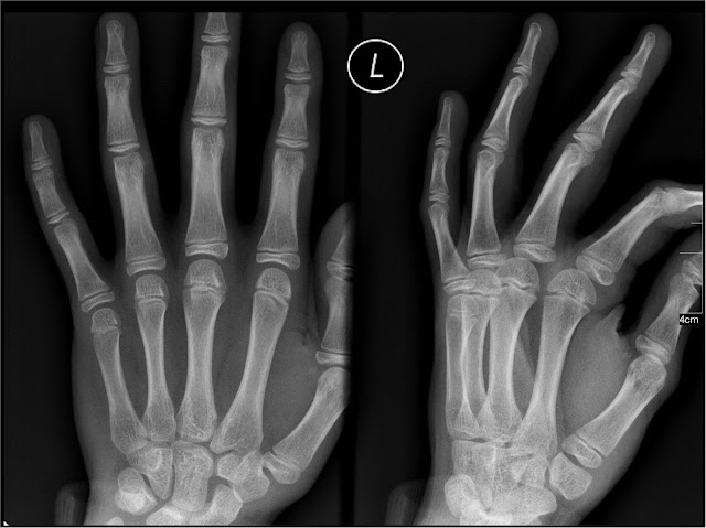
The appearance of different body tissues on an X-ray picture depends on how much each tissue blocks the passage of the radiation. Bone, which blocks the most radiation, appears white, while air, which blocks the least, appears black. Tissues other than bone are represented by various shades of grey.

Digital X-ray Imaging

Traditionally, medical X-ray images have been exposed onto photographic film. X-ray films require processing before they can be viewed, however, and take up large amounts of storage space in hospitals and doctors’ offices. Over recent years, digital X-rays have become increasingly popular for radiological imaging.

To obtain a digital X-ray picture, an electronic detector is used in place of a photographic film. This detector sends the signal produced by the X-rays to a computer, which converts the signal to a digital image that can be viewed on screen immediately and saved as required.

Radiological Imaging Using Medical X-rays

X-rays are especially useful for imaging the bones, but soft tissues such as the lungs and digestive system can also be viewed and X-ray images are important in the diagnosis of illnesses such as lung cancer and kidney stones.

Other applications of medical X-rays include:
  • dental X-rays, which can detect problems with the teeth at an early stage
  • mammography to screen for breast cancer (digital mammography and computer-aided detection of abnormalities in the breast are recent advances in this field)
  • fluoroscopy, which produces moving images used to examine the action of the gut or to guide a procedure such as implantation of a cardiac pacemaker.

Are Medical X-rays Safe?

As a type of radiation, any exposure to X-rays carries a risk of cancer. However, the amount of radiation produced by a single X-ray picture is extremely low compared with the background radiation that a person experiences every day from natural sources such as cosmic rays and radioactive minerals in the earth. Health professionals consider the benefits of an accurate diagnosis and treatment to far outweigh the small risk involved in X-ray imaging.

The risks of X-rays are greater for young children and unborn babies, but the doctor will always bear this in mind when deciding on the need for medical imaging.
Other Uses for X-rays in Medicine

X-rays are used to treat disease in the form of radiotherapy, while CT scanners employ multiple X-ray beams to produce detailed pictures of soft tissues as well as bones. However, a CT scan exposes the patient to more radiation than a conventional X-ray film or digital X-ray.

buy steroids legally uk

Popular posts from this blog

Common Signs of Heat Stroke

According to MedicineNet in "Definition of Heat Stroke," heat stroke occurs when the body's core temperature rises above 104°F (40°C) and is accompanied by hot, dry skin and central nervous system abnormalities such as delirium, convulsions or coma. Since heat stroke is a life-threatening condition, it is important to watch people for heat stroke symptoms. MedicineNet states that heat stroke that results from exposure to high environmental temperatures is termed nonexertional heat stroke, and heat stroke that results from strenuous exercise is called exertional heat stroke. Regardless of the type of heat stroke a person experiences, heat stroke signs are life-threatening, and immediate medical attention is necessary when heat stroke symptoms begin. When the body cannot control its temperature, heat stroke occurs because the sweating mechanism fails. The body is not able to cool down. Body temperature can rise to 106°F (41.1°C) or higher within a few minutes. Hea

Causes of Easy Bruising: Reasons Why People Bruise Easily

Bruising , a reddish or purple discoloration under the skin, most often results from trauma to the small blood vessels, called capillaries, but can also occur spontaneously. How and Why Bruises Occur Blood leaks out of the capillaries and accumulates under the skin, gradually absorbing over several days. Bruising most often occurs because people run into objects or experience other trauma. Most bruising is easily explained, but frequent bruising that occurs without obvious cause needs prompt investigation, since several serious diseases can cause bruising. In general, women bruise more easily than men. How Aging Increases the Risk of Easy Bruising Bruising increases as people age for several reasons. Skin thins as people age and the capillaries become more fragile. The layer of fat that cushions blood vessels and protects them from injury becomes thinner as well. Older people often take medications and supplements that thin the blood and contribute to easy bruising. Visi

Stye - Symptoms and Treatment

A stye is an infection, typically a bacterial infection, which causes a painful red lump either on the edge or inside of the eyelid. Bacteria grow at the root of an eyelash follicle or inside an oil gland. The bacteria can be a result of poor hygiene, touching the eyes with unwashed hands or chronic inflammation. The stye resembles a pimple or a boil and is usually filled with pus. A stye does not pose a risk to vision and most often heals without treatment within a week. However, a stye may require treatment with a doctor if the infection does not resolve with at-home remedies. Symptoms of a Stye A stye is not usually hard to spot. Not only does a red lump form on either the top or bottom eyelid, but pain is also an immediate symptom of the condition. Typical symptoms of a stye include: red lump on the eyelid similar to a pimple watering of the eye eyelid pain and swelling clear or yellow fluid collecting in the stye A normal stye will come to a head in approximately three to