Olser-Weber-Rendu syndrome (OWR), also known as Hereditary Hemorrhagic Telangiectasia (HHT), is an inherited condition affecting the blood vessels. It affects approximately one person out of 10,000, or about 1.2 million people throughout the world.
OWR syndrome has an autosomal dominant pattern of inheritance, meaning only one copy of the abnormal gene is necessary to cause the disease and to pass it on. Each child of an individual who has the disorder has a 50% chance of inheriting it. The vast majority of those affected have a family history of the disorder.
Treatment varies depending on the severity of the condition. Mild cases may require little or no treatment. Treatment for more severe symptoms may include iron supplements for anemia or laser therapy to seal telangiectasias. Chronic bleeding from the GI tract may require endoscopy and treatment by laser therapy or cauterization of AVMs. Pulmonary AVMs may be treated with embolization, which is insertion of a tube through a vein in the groin area that is used to place a balloon in the lung to block the bleeding artery. Brain AVMs may be treated by surgery, embolization, or stereotactic radiosurgery, which uses a focused beam of radiation.
Other treatments include hormone therapy with estrogen, or aminocaprioic acid, which improves clotting. In cases of severe blood loss, blood transfusions may be necessary. Many people who have Osler-Weber-Rendu syndrome do not have severe symptoms, and require minimal treatment to manage the condition, but early screening and proper diagnosis are crucial.
Causes of Osler-Weber-Rendu Syndrome
Olser-Weber-Rendu syndrome is a genetic disorder caused by an abnormality in either the endoglin gene (ENG) on chromosome 9, or the activin receptor-like kinase 1 gene (ALK1) on chromosome 12. Both of these genes are involved in blood vessel formation. A mutation in either of these genes will result in similar OWR symptoms, and those who have the disorder generally only have an abnormality in one of the genes.OWR syndrome has an autosomal dominant pattern of inheritance, meaning only one copy of the abnormal gene is necessary to cause the disease and to pass it on. Each child of an individual who has the disorder has a 50% chance of inheriting it. The vast majority of those affected have a family history of the disorder.
Symptoms of Osler-Weber-Rendu Syndrome
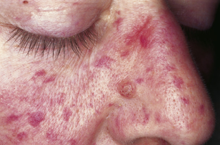
Most symptoms of OWR syndrome are due to abnormal formation of capillaries, tiny vessels that normally connect arteries to veins. Abnormalities in capillary formation cause defects known as arteriovenous malformations (AVMs), fragile areas in the vessels that can easily rupture. AVMs may occur on the surface of the skin or in the lungs, brain, liver, stomach or gastrointestinal tract. Symptoms often begin to appear in affected people when they are between ten and twenty years old, and increase with age. Individuals with OWR will not necessarily have all of these symptoms.- Telangiectasias are small AVMs that may appear on the skin as red spots on the face, hands, lips, or inside the mouth. They may bleed spontaneously or from minor trauma.
- Nosebleeds (epistaxis) usually begin to appear around 12 years of age, and are due to telangiectasias in the nose.
- Anemia can be caused by blood loss from frequent nosebleeds or bleeding from AVMs elsewhere in the body.
- Pulmonary AVMs cause bleeding in the lungs, increase the risk of bacterial infections by interfering with normal filtering processes, and cause low blood oxygen levels, migraine headaches, and can possibly lead to stroke.
- Brain AVMs can cause headaches, seizures, paralysis or stroke.
- Liver AVMs can interfere with normal circulation of the blood and lead to increased risk of heart failure.
- Gastrointestinal AVMs can cause significant loss of blood, leading to anemia.
Diagnosis and Treatment of Osler-Weber-Rendu Syndrome
OWR syndrome is usually diagnosed by observation of symptoms such as frequent nosebleeds and telangiectasias, and whether there is a family history of the disorder. Blood tests can detect anemia and monitor blood oxygen levels; chest x-rays or EKGs can assess if lungs and heart are normal; ultrasound is used to find AVMs in the stomach or liver; MRIs are used to look for AVMs in the brain. Gastrointestinal bleeding can be detected by stool samples.Treatment varies depending on the severity of the condition. Mild cases may require little or no treatment. Treatment for more severe symptoms may include iron supplements for anemia or laser therapy to seal telangiectasias. Chronic bleeding from the GI tract may require endoscopy and treatment by laser therapy or cauterization of AVMs. Pulmonary AVMs may be treated with embolization, which is insertion of a tube through a vein in the groin area that is used to place a balloon in the lung to block the bleeding artery. Brain AVMs may be treated by surgery, embolization, or stereotactic radiosurgery, which uses a focused beam of radiation.
Other treatments include hormone therapy with estrogen, or aminocaprioic acid, which improves clotting. In cases of severe blood loss, blood transfusions may be necessary. Many people who have Osler-Weber-Rendu syndrome do not have severe symptoms, and require minimal treatment to manage the condition, but early screening and proper diagnosis are crucial.
